Functional electronic stimulation (FES) uses the body’s existing nerves and muscles to create a desired contraction in the targeted muscle. Patients who have had an upper motor neurone injury, caused by stroke or cerebral palsy, can usually benefit.
FES works by sending an electrical impulse, either along a nerve that is located close to the surface of the skin, or through a muscle. When the impulse is passed through the muscle it causes a contraction. The big advantage of FES is that it uses the body’s existing muscular system to cause movement and control it; this means that an individual’s muscle strength can be increased or, at least, maintained.
There are also possible benefits to the sensory system. There is some evidence to suggest that FES can assist remyelination in conditions such as multiple sclerosis, or that it improves control by sending signals to the brain. Research is being carried out to validate this.
Above: Patient being assessed upon a pressure sensor within LOC's Gait Lab
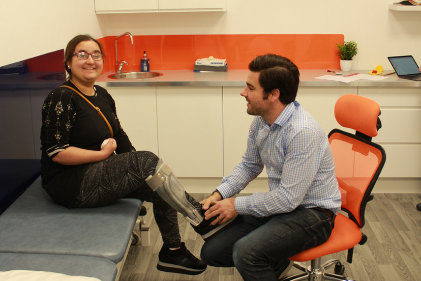
At the initial consultation, an in-depth history of a patient’s neurological condition will be established. Range of movement, alignment and existing muscle power will be analysed while video gait analysis will study any areas of weakness in slow motion.
If a LOC clinician considers that an individual’s condition is appropriate for FES, then we will test the MyGait system. This involves applying electrodes to targeted areas of the body in order to create the desired muscle contractions. As anatomy can vary between individuals finding the perfect position for the electrodes can take some time. Your clinician will then test the signal from the electrodes and fine-tune the type of muscle contraction that is required.
Once that has been successful, your clinician will use the system while you are walking. MyGait has very sophisticated software that allows fine-tuning of both the timing and strength of the impulses wirelessly as the patient walks. It is possible to see immediate and remarkable improvements.
We have the following facilities and amenities at our Kingston Upon Thames location:
We also have the Gait Laboratory for orthotics patients and Onsite Manufacturing for speedy turnarounds and adjustments whilst you wait.
An insole is a contoured orthotic device which alters the characteristics and biomechanics of the foot and ankle area. Biomechanics are concerned with mechanical laws and how they affect the living body, especially the musculoskeletal system.
They are removable devices, often made from plastic, that are designed to fit inside a shoe to provide additional support for your feet. As well as offering shock absorption, an insole can help distribute the weight of your body more effectively across the foot and can be made bespoke to cover a range of biomechanical conditions.
If you have symptoms in your feet, ankles, hips or your lower back that are intermittent or were not there to start with in early life, and have started to cause you pain over a period of time, bespoke orthotic insoles could be an excellent option.
If you have already tried rest, icing, compression and elevation and your feet have not recovered, we recommend a biomechanical assessment to consider the possibility of insoles. They are a non-invasive approach to treatment and in many cases, are a great option for symptoms that are not severe enough to warrant surgical intervention. Alternatively, they can be considered as an option prior to surgery.
We will send patients away when an insole is not appropriate, if a patient is suffering with iliotibial band syndrome for example, the problem can be helped with physiotherapy and a stretching programme. That’s what our biomechanical assessment is all about; determining whether there would be any benefit from altering the alignment of your feet.

See how a thorough gait analysis and a correctly-fitted, bespoke Reciprocating Gait Orthosis (RGO) helped Ted, a spinal surgery and cancer survivor, improve his rehabilitation and mobility goals, getting him back on his feet again.

We are proud to announce the launch of our latest innovation in non-surgical treatment for pectus deformities. Our new dynamic chest compressor is one of the slimmest pectus braces on the market and is designed to reshape the chest without the need for invasive surgery.

Rosie’s very severe plagiocephaly was no problem for the LOCBand Lite 3D-printed cranial remoulding helmet, going from 16mm to 2mm in just six months.

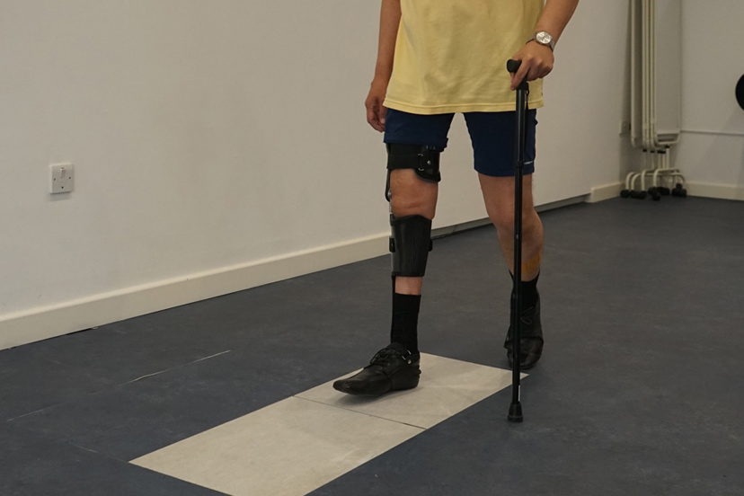
When John came to see us, his ankle was in a bad way. He had around 60mm of his tibia missing and not much if any talus present. He needed crutches to support him to walk. A gait analysis and a new bespoke carbon fibre knee ankle foot orthosis (KAFO) later and he is able to walk again without crutches.

Matilde travelled from Chile to LOC for bracing treatment for her adolescent idiopathic scoliosis. Now, nearly a year and a half since she started wearing her brace, she has achieved near-total correction of the curvature of her spine. This is her scoliosis bracing story.

After only 6 months of wearing bespoke pectus braces from The London Orthotic Consultancy, Will started to notice a visible difference in his pectus carinatum.

After trying out several scoliosis braces in Romania, Ukraine and Turkey, Iulia begins treatment with the LOC Scoliosis Brace and is already seeing results in a matter of months. Here her mum, Raluca, describes how and why they came to LOC for her treatment.

Through bracing treatment with the dynamic chest compressor, Jack has achieved 90% correction in his pectus carinatum after only two months. Here, mum describes Jack's non-surgical treatment journey.